

Scanning Electron Microscope
Scanning Electron microscopy (SEM) uses a highly focuses electron beam which can be scanned in a raster on the sample surface. The intensity of secondary electrons produced at each point is used to form a picture of the sample. Using backscatter electron (BSE) detector materials with different composition as different (greyscale) contrast can be imaged. Using EDX detector; enegy dispersive x-ray spectroscopy (EDS) for analyzing main components and low-level contaminants in samples and elemental distribution maps to show the distribution on the sample of an element of interest can be performed.
Quanta 250 SEM enables characterization of non-condactive and without coating and ESEM capability enables charge-free imaging of hydrated specimens.
The electron microscopes in the center and their detectors is as follows:
FEI QUANTA 250 FEG:
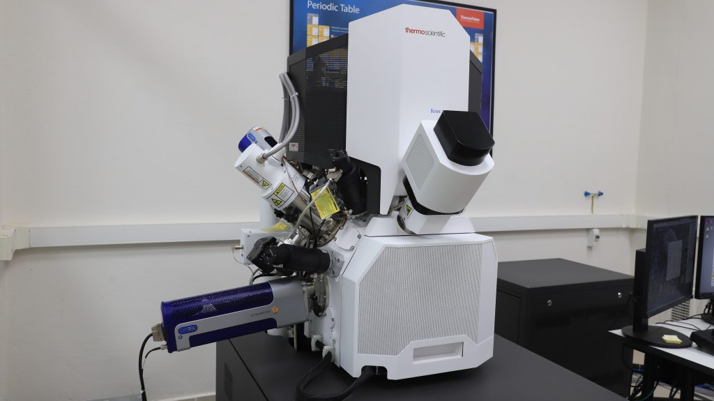
SCIOS2 DUAL BEAM (FIB)
ZEISS EVO10